Here, how to output area, length, and width information, as well as the average pixel value for each organ from images using ImageJ is introduced. For example, leaf area is needed to calculate the Leaf Area Index (LAI), the area of leaves per unit land area, which is an important parameter for crop dry matter production. Image pixel values can be used to compare leaf colors relatively.
ここでは、ImageJを用いて画像から各器官の面積、長さ、幅の情報と、平均画素値を出力する方法を紹介します。例えば、葉の面積は作物の乾物生産に重要なパラメータである単位土地面積当たりの葉の面積(Leaf Area Index: LAI)の算出に用いたり、画像の画素値は葉の色を相対的に比較するのに用いたりすることができます。
- Calculation of area, length, and width and preparation for pixel value measurement
面積、長さ、幅の計算と画素値測定の準備- File -> Open
you can open an image file to process
処理する画像ファイルを開く。 - Crop image
画像の切り出し - Set scale
スケールの設定- put a straight line along with the scale
スケールに沿って直線を設置

- Analyze -> Set Scale
- Known distance: Actual length of the set straight line
設置した直線の実際の長さ - Unit of length: Unit of length of actual length ex) cm, m
設置した直線の実際の長さの単位(例:cm, m)
- Known distance: Actual length of the set straight line
- put a straight line along with the scale
- Extract region to process
処理する器官の領域を抽出する- Image -> Type -> Lab Stack
Conduct Lab stack
Lab色空間に変換する - move to a channel
処理を行うチャンネルに移動- leaf -> "a" channel

- stem-> "b" channel
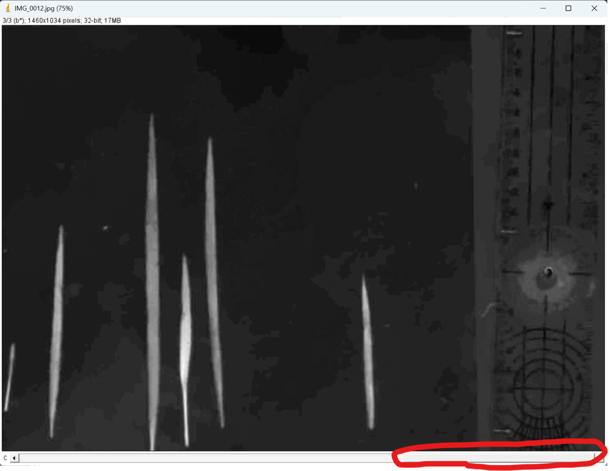
- leaf -> "a" channel
- Image -> Adjust -> Threshold
- Please find a method to extract regions precisely
正確に各器官の領域を抽出できるアルゴリズムを選択
Selection area can be inverted by put check in Dark background check box
"Dark background"にチェックを入れることで、選択領域を反転させることができる(If you want to set the threshold manually, move the bar below to set the threshold, then click "Set")
(もしマニュアルで閾値を設定したい場合は、以下のバーを動かして閾値を設定した後に、"Set"をクリックする)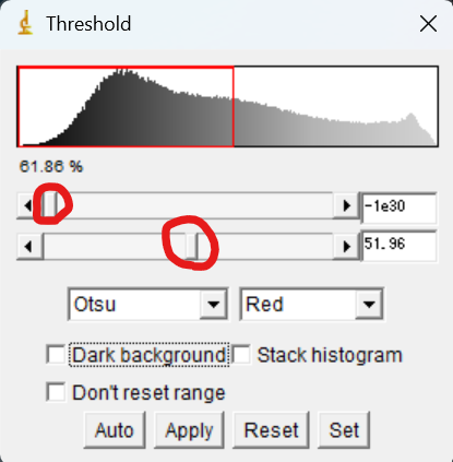
Then, click "Apply" to put threshold
"Apply"をクリックする- "Convert to Mask"

- Method: Selected algorithm
Backgroud: Light
☑ Calculate threshold for each image
(If you have set a manual threshold, uncheck it)
(もしマニュアルで閾値を設定していた場合はチェックを外す)
Then, click "OK"
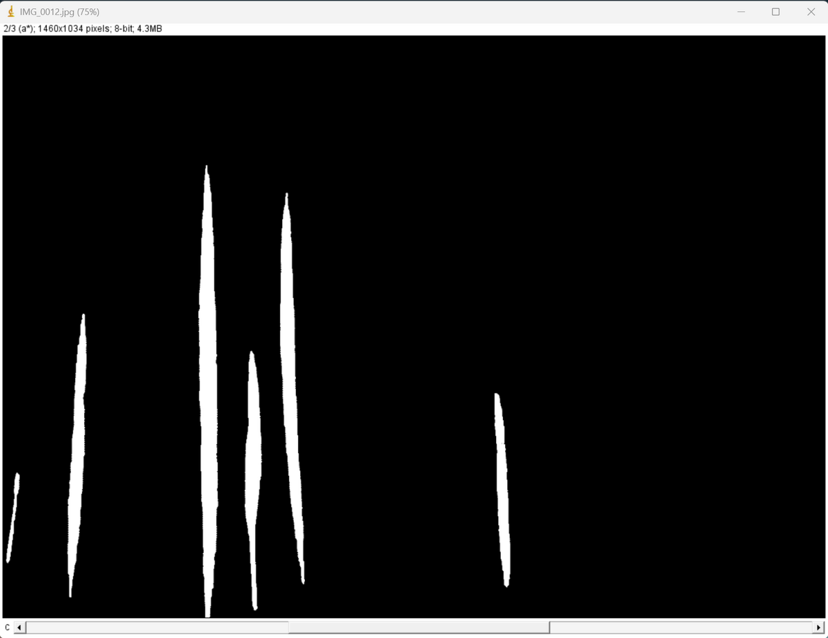
- You can get a binary image
here you can see extracted six leaves
このように、2値化された画像が出力される
この例では、6つの葉が抽出されます
- Image -> Type -> Lab Stack
Conduct Lab stack
- Analyze -> Set Measurements
Set parameters to be measured
計測するパラメータの設定- For more information, the following is helpful.
詳細は以下のサイトが参考になります。
imageJで測れるものは?Set Measurementsの設定 (life-science-project.com)
- For more information, the following is helpful.
- Analyze -> Analyze Particles
Perform measurements of each organ
各器官の計測の実施- Size: Minimum and maximum size of each part to be analyzed. By increasing the minimum size, small noise particles can be removed for analysis.
解析対象となる各器官の最小サイズと最大サイズです。最小サイズを大きくすることで、小さなノイズ粒子を取り除いて解析することができます。 - Put check in following four check boxes
Click "OK"
以下の4つのチェックボックスにチェックを入れてください。
"OK "をクリック
- Click "No"
"No "をクリック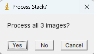
- Size: Minimum and maximum size of each part to be analyzed. By increasing the minimum size, small noise particles can be removed for analysis.
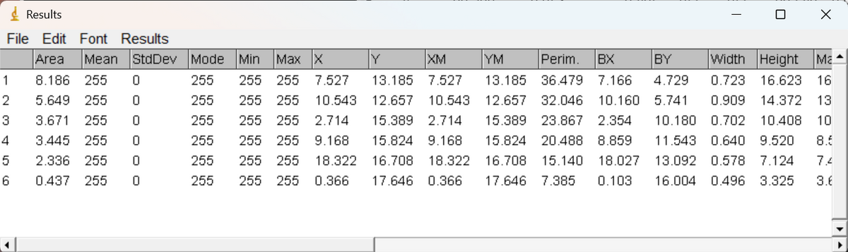
- You can get three windows
3つのウィンドウが開きます。- "Results"
Here, you can see the area, width, height, and so on for each leaf.
"Results"
ここでは、各葉の面積、幅、高さなどを確認することができます。
- "Summary"
Here, you can see the total area, average size and so on.
"Summary"
ここでは、総面積や平均サイズなどを確認することができます。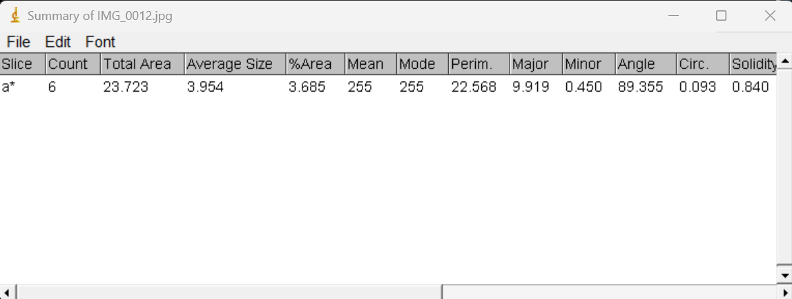
- File -> Save As
You can save these tables as csv file
これらのテーブルは、csvファイルとして保存することができます。 - "ROI Manager"
ROI is the information of region of each part
Select all the ROIs, then
ROI(region of interest)とは、各器官の各領域の情報です。すべてのROIを選択し、
More -> Save
You can save the ROI information as zip file
ROI情報をZIP形式で保存することができます
- "Results"
- File -> Open
- Calculation of the average pixel value for each part
各器官の平均画素値の算出- File -> Open
You can open the cropped image file which you saved
保存した切り抜き画像ファイルを開く - Perform set scale as before
- File -> Open
You can open the zip file of ROI which you saved
保存したROIのZIPファイルを開く - This time we will process the red channel of the RGB image.
今回はRGB画像の赤のチャンネルを処理してみます。 - Image -> Type -> RGB Stack
- Select a channel to process
This is an example of red channel
処理するチャンネルを選択する
これは赤色のチャンネルの例です
- Select all ROIs and click "Measure"
すべてのROIを選択し、"Measure "をクリック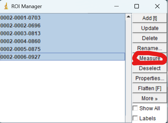
- You can get the results of the mean pixel value of red for each leaf
各器官の赤色の平均画素値の結果を取得することができます
- File -> Open
You can open the cropped image file which you saved
- It is a good idea to use acrylic plates like this one to keep the leaves open and fixed in less reflective conditions.
反射の少ない条件で葉を開いて固定した状態で撮影を行うため、このようなアクリル板を使用するのがよいでしょう。
【楽天市場】ノングレア アクリル板 A3サイズ(297mm×420mm)反射低減 透明 3mm厚 角丸 糸面取り 側面磨き 付き:看板ショップ (rakuten.co.jp)